
X-Rays Durban
X-Rays are a type of electromagnetic radiation that are commonly used in medical imaging to create images of the body’s internal structures. X-Ray imaging works by passing high-energy X-Ray photons through the body, with denser materials such as bones or tumours absorbing more of the radiation than surrounding tissues. The resulting image shows the shadow patterns created as the X-Rays pass through the body, producing a two-dimensional representation of the internal structures.
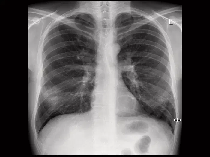
General X-Rays
X-Rays can be used to diagnose a variety of medical conditions, including broken bones, pneumonia, dental problems, and tumours.
They are generally safe and painless, but can carry some risks thus a referral letter is required from your treating practitioner.
Images are stored digitally to enable access to these studies by doctors, facilitating comparisons and diagnoses.
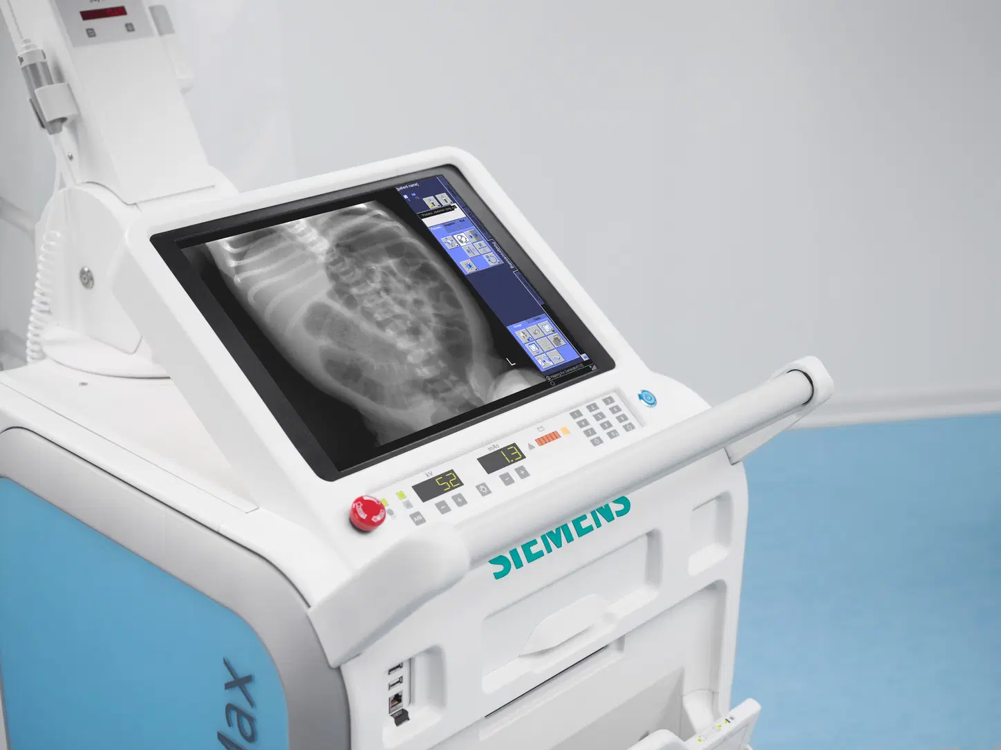
Portable X-rays
A portable x-ray can conveniently be taken to the patient in a hospital bed or the emergency room.
The x-ray tube is connected to a flexible arm that is extended over the patient while the x-ray film holder or image recording plate is placed beneath the patient.